Subcellular Mitochondria Structure Prediction in Label-free Microscope Images Using Convolutional Neural Networks
Abstract
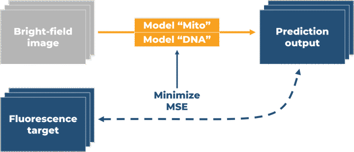
Fluorescence microscopy enables us to analyze the subcellular structure of the living cell with specific labeling, but it also comes with potential problems of phototoxicity. In this study, we adopt the published label-free method by Allen Institute to train and predict 3D fluorescence images from our transmitted light microscopy images of cardiac myocyte-derived cell line AC16.
Type
Publication
In * The 20th International Conference on Systems Biology*
Chan-Min Hsu
Research Assistant
My research focus on biomedical image segmentation using deep learning techniques.